In an article published in the journal Nature, researchers assessed the clinical utility and reliability of an automated eyelid measurement system, using neural network (NN) technology. The study, involving 300 subjects with Graves’ orbitopathy (GO), ptosis, and controls, developed an NN-based system to measure eyelid parameters. Results demonstrated high agreement with manual measurements, indicating the system's accuracy.
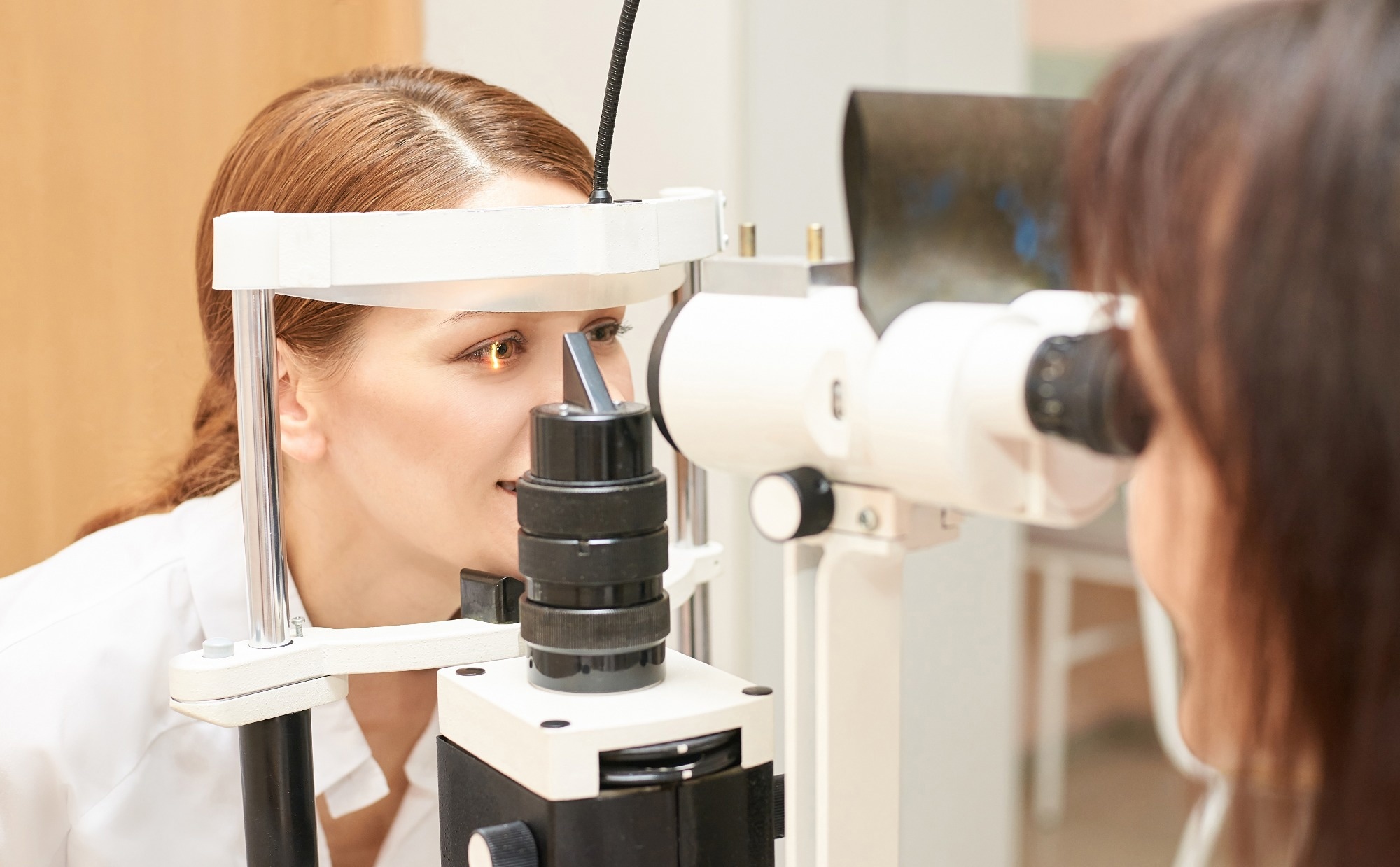
 Study: Neural Network-Based Automated Measurement System for Eyelid Assessment. Image credit: elenavolf/Shutterstock
Background
Eyelid abnormalities are common in ophthalmic disorders like GO, ptosis, and orbital tumors, necessitating a thorough evaluation of eyelid morphology. Current metrics, such as margin-reflex distance one (MRD1) and margin-reflex distance two (MRD2), palpebral fissure height (PFH), and eyelid length, lack consistency and reproducibility in manual measurements. Additionally, these metrics often struggle to capture the curved aspects of eyelids. Attempts to automate eyelid morphology evaluation have been made, but semi-automatic methods and low-contrast transitions in images pose challenges.
This study addressed these challenges by introducing an NN-based automated eyelid measurement system. Unlike manual and semi-automatic techniques, NNs provided consistent measurements through automated segmentation, overcoming issues related to image contrast. Leveraging deep learning techniques, researchers explored the feasibility of NNs in accurately establishing reference lines, such as the intercanthal distance, for measuring the curved eyelid contour. The authors aimed to fill existing gaps by evaluating the clinical utility and reliability of this NN-based system and comparing it with traditional methods. The proposed approach not only sought to automate measurements but also promised to effectively detect eyelid abnormalities.
Method
The research, approved by the institutional review board of Chung-Ang University Hospital, aimed to assess the clinical utility and reliability of an NN-based automated eyelid measurement system. The retrospective analysis included 300 subjects with normal eyelids, GO-related eyelid retraction, and eyelid ptosis. Facial photographs were taken using standardized conditions, and manual measurements of parameters like MRD1, MRD2, and upper and lower eyelid lengths were performed using ImageJ software.
An automated system based on NNs was developed to measure the same parameters, utilizing image segmentation and proportional equations. Additionally, the system divided the eyelid margin into partitions for automated detection of abnormalities. The authors introduced the concept of the intercanthal distance as a reference line for improved accuracy in measuring curved eyelid contours. The automated system effectively detected abnormalities with a minimum of 24 partitions.
Deep learning techniques, specifically the DeepLab V3+ model, were employed for image segmentation, demonstrating good performance with a mean intersection over union (mIoU) metric. Statistical analyses included analysis of variance (ANOVA), Wilcoxon rank-sum test, paired t-test, and calculation of intraclass correlation coefficients (ICC) for agreement assessment. Bland–Altman plots visualized discrepancies between manual and automated measurements.
The NN-based automated system showcased clinical usefulness and reliability, offering precise measurements and effective detection of eyelid abnormalities. The authors provided a comprehensive methodology combining image segmentation, neural networks, and statistical analyses for accurate and consistent eyelid assessment.
Results
The research involved 300 subjects (mean age 50.5 ± 18.6 years), categorized into normal, ptosis, and GO groups. The automated eyelid measurement system, based on NNs, exhibited consistently higher values for MRD1 and MRD2, upper eyelid length, and lower eyelid length compared to manual measurements. Despite slight variations, there was good overall agreement between manual and automated measurements, with ICC indicating excellent correlation for MRD1 and MRD2 (0.972 and 0.937, respectively).
Bland–Altman plots illustrated narrow limits of agreement, signifying high consistency. Subgroup analysis revealed consistently strong ICC values for MRD1 and MRD2, while upper and lower eyelid lengths exhibited fair to good agreement. The system's performance in detecting abnormal eyelid segments improved with 24 partitions (15-degree intervals), balancing simplicity and accuracy. The researchers demonstrated the clinical utility and reliability of the automated system, offering precise measurements and effective detection of eyelid abnormalities, especially in MRD1 and MRD2.
Discussion
An automated eyelid measurement system based on NNs was developed to quantitatively measure eyelid parameters, including MRD1 and MRD2. The system demonstrated high accuracy and efficiency compared to manual measurements, addressing interobserver variability and time consumption associated with conventional methods. While the automated measurements showed slightly higher values than manual ones, particularly in patients with ptosis or GO, the overall agreement was robust.
The system effectively detected abnormalities in the eyelids using 24 partitions. Despite the promising results, the authors acknowledged limitations such as a relatively small sample size and the need for additional research to refine pupil center estimation for cases with negative MRD1 values. Overall, the NN-based system held the potential for clinical applications, either as a standalone application or integrated into existing medical devices, paving the way for future studies to explore broader applications and clinical utility.
Conclusion
In conclusion, the researchers introduced an automated eyelid measurement system utilizing NNs, demonstrating high accuracy and efficiency in comparison to manual methods. Involving 300 subjects with diverse eyelid conditions, the NN-based system offered precise measurements of parameters like MRD1 and MRD2, effectively detecting abnormalities.
The authors proposed an innovative approach, measuring the intercanthal distance for enhanced objectivity. Despite slightly elevated values in automated measurements, particularly in specific conditions, the overall agreement remained robust. The system's clinical utility and reliability suggested its potential integration into medical practice, promising comprehensive and accurate eyelid assessments.
Journal reference:
- Nam, Y., Song, T., Lee, J., & Lee, J. K. (2024). Development of a neural network-based automated eyelid measurement system. Scientific Reports, 14(1), 1202. https://doi.org/10.1038/s41598-024-51838-6, https://www.nature.com/articles/s41598-024-51838-6