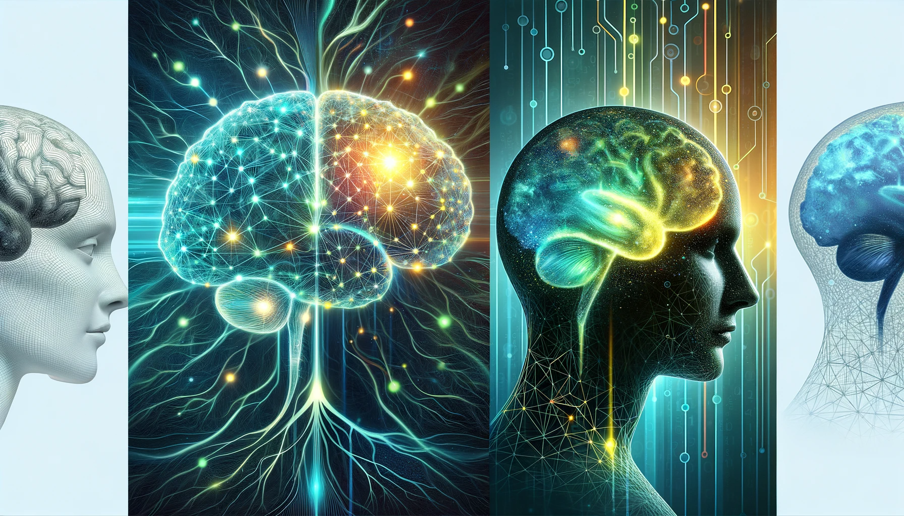
A recent publication in the journal Nature explored the use of machine learning to simulate the human brain and discussed challenges in gathering comprehensive data, emphasizing the need for collaboration, and defining modeling targets.
 Study: Revolutionizing Neuroscience: Machine Learning's Role in Simulating the Human Brain. Image credit: Generated using DALL.E.3
Background
In the pursuit of whether a computer can be programmed to simulate the human brain, a question that has intrigued mathematicians, theoreticians, and experimentalists, lies a profound inquiry. It is driven by the ambition to create artificial intelligence (AI) and the notion that understanding a complex system such as the brain necessitates the mathematical or computational reproduction of its intricate behaviors. This journey of exploration commenced in the 1940s with the development of simplified models of brain neural networks, which were deeply rooted in early biological inspirations.
Nonetheless, the current trajectory raises an altered question: Could machine learning be leveraged to construct computational models that faithfully simulate the activities of brains? Starting in the 1970s and intensifying since the mid-2000s, neuroscientists have meticulously crafted connectomes—comprehensive maps detailing the connectivity and morphology of neurons, portraying a static snapshot of the brain at a specific moment. Concurrently, advancements in functional recordings have enabled the observation of neural activity at the single-cell level over time.
As of now, the integration of these disparate data sources or their simultaneous acquisition from a single specimen's entire brain remains relatively unexplored. Nevertheless, the growing depth, scale, and diversity of these datasets, especially in simpler model organisms, are fostering a novel approach to brain modeling. This approach entails training AI programs on connectome data to replicate the expected neural activity in biological systems. Nonetheless, computational neuroscientists and their cohorts must grapple with several challenges before fully embracing machine learning in their quest to simulate entire brains.
Advancements in brain mapping
The journey of brain mapping commenced nearly half a century ago, epitomized by the painstaking 15-year endeavor in the roundworm Caenorhabditis elegans. Over the past two decades, automated tissue sectioning and imaging have revolutionized the acquisition of anatomical data, while computational and automated image analysis have streamlined data interpretation.
Connectomes now exist for the entire brain of C. elegans, larval and adult Drosophila melanogaster flies, and select segments of the mouse and human brain. Despite their advancements, these anatomical maps possess notable gaps. Mapping electrical connections, in conjunction with chemical synaptic ones, remains a formidable challenge. Neuronal focus has somewhat overshadowed the role of non-neuronal glial cells in information processing. Additionally, many aspects of gene expression and protein presence within mapped cells remain enigmatic.
Nevertheless, these maps have already begun yielding insights, unraveling the mechanisms behind behaviors such as aggression and the neural circuits governing spatial awareness in D. melanogaster. Efforts to map the entire mouse brain are underway, although it may take a decade or more given its immense complexity.
Machine learning for advancing brain simulation and understanding
In tandem with the progress in connectomics, scientists have made substantial headway in capturing gene expression patterns and neural activity across entire vertebrate brains. Mathematical models driven by physics have historically dominated attempts to simulate brain activity, leveraging mathematical descriptions of real neurons and unverified assumptions about network connectivity. While anatomical maps have provided valuable guidance, they have also exposed the limitations of an anatomy-driven approach. Machine learning emerges as a promising avenue to address these challenges.
By optimizing thousands or even billions of parameters with guidance from connectomics and other data sources, machine learning models can mimic the behavior of real neural networks, as observed through cellular-resolution functional recordings. These models can combine conventional brain modeling techniques with empirically optimized parameters or adopt 'black box' architectures devoid of explicit biological knowledge. This approach can enhance the rigor of modeling brain networks comprising thousands or more neurons, enabling comparisons between simpler, computationally efficient models and more complex ones incorporating detailed biophysical information.
While challenges and limitations persist in adopting machine learning for brain modeling, this approach holds promise. It can potentially fill gaps in existing datasets and reduce the need for increasingly detailed measurements of individual biological components. As comprehensive data becomes more abundant, it can be seamlessly integrated into these models, ushering in a new era of brain simulation and understanding.
Overcoming challenges in brain simulation with machine learning
Emulating the brain through machine learning presents data quality challenges, requiring comprehensive data collection from entire brains or organisms. Simple nervous systems may assume connectivity consistency, but complex ones necessitate training on specimen-specific data due to connectome variability.
Attaining multimodal data demands collaboration, investment, and agreed-upon metrics. Combining traditional neural networks with machine learning can replicate sensory processing in the brain, offering analytical convenience. The key question is whether existing brain mapping data can effectively train machine-learning models to replicate biological neural activity, with potential failures signaling a need for more extensive mapping.
In summary, the current study explored the potential of machine learning to enhance our understanding of the brain. It highlights the challenges and promise of using machine learning to simulate and study brain activity.